

Walking into the “Body Worlds” exhibit at the John P. McGovern Museum of Health and Medical Science in the heart of the Houston museum district is an intriguing experience. Most people tend to know the basic parts that make up our anatomy—whether it’s the bones, muscles, or nerves. However, being able to see face-to-face plastinated bodies exposes a whole new world of our bodies that we call the body. Plastination is a body preservation technique developed by Gunther von Hagens in which the body is coated with resin.
“Body Worlds” is a traveling exhibit that is shown across Europe and North America, with most of its permanent locations being in European cities such as Berlin and Amsterdam. In Houston, the exhibit consists of displays of fully plastinated bodies, organs and bodily systems, revealing the inner anatomy of a human in extreme detail. However, it’s not permanent. The exhibit returned to Houston on Feb. 5 and leaves on Oct. 5.
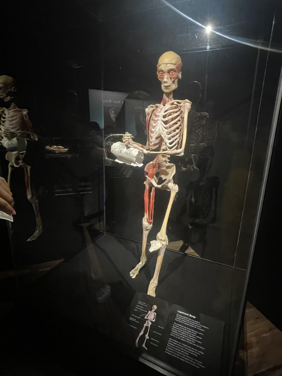
The first site greeting visitors by the entrance of the exhibit is a full-body plastinate titled “Ligament Body.” The plastinate shows how the ligaments come together to support the skeletal system. Positioned next to the plastinate’s glass display is a plaque that gives a short but in-depth description of what the ligament body is alongside a diagram labelling various parts such as the patella, tibia and the temporal muscle.
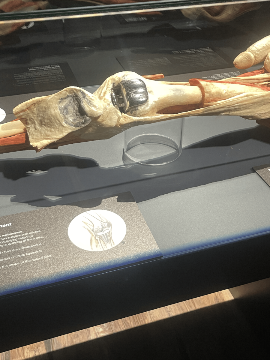
Moving on, the next section concerned the different joints in the body, such as the joints in the knees, what happens after they are broken and how to seek treatment to ail those broken joints. Reading through the description, it defines the joint as “the point at which two bones meet.” Right next to that model of the knee joint displays a model of a stainless steel knee replacement. It shows the knee replacement being lodged into the same position of a knee, hence the name knee replacement. After that, a model of a broken femur is shown with a large stainless steel rod implanted into it with the plaque, describing the process of healing that happens after someone breaks a bone in their body.
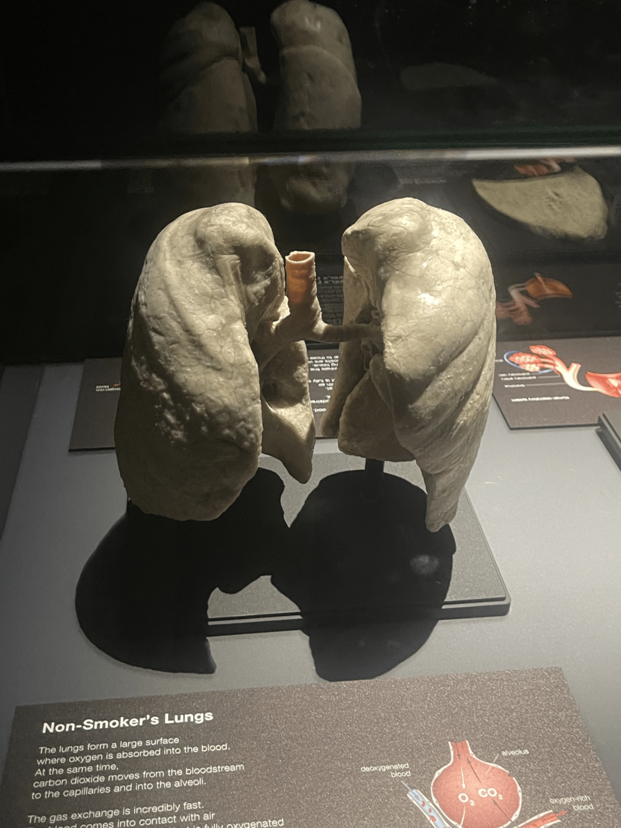
The next section of the exhibit discussed the respiratory system featuring parts such as the mouth, the trachea and the most important part of the system — the lungs. This part of the exhibit details damage that can be done to the respiratory system from smoking. The exhibit shows a side by side diagram of a healthy non-smoker’s lung next to a smoker’s lung, with a plaque warning viewers about the dangers of smoking.
Whether it was teaching viewers about the damage that can be done to your body or just informing about the human body, visiting the “Body Worlds” exhibit at the Health Museum was a once in a lifetime experience. Being able to experience this temporary exhibit with all the physical models was genuinely interesting and generally immersive for viewers into a world that was familiar but also new. With the exhibit doing its job in peaking curiosity, the Health Museum excels in its ability to show the human body in a light that most haven’t seen it in.